Chemotactic Bacteria Facilitate the Dispersion of Nonmotile Bacteria through Micrometer-Sized Pores in Engineered Porous Media
María Balseiro-Romero et al.
Abstract
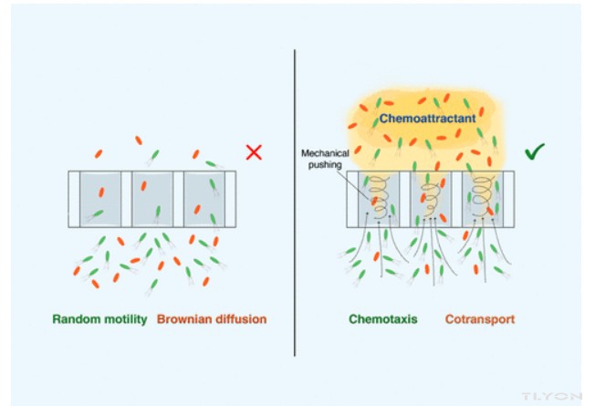
Recent research has demonstrated that chemotactic bacteria can disperse inside microsized pores while traveling toward favorable conditions. Microbe–microbe cotransport might enable nonmotile bacteria to be carried with motile partners to enhance their dispersion and reduce their deposition in porous systems. The aim of this study was to demonstrate the enhancement in the dispersion of nonmotile bacteria (Mycobacterium gilvum VM552, a polycyclic aromatic hydrocarbon-degrader, and Sphingobium sp. D4, a hexachlorocyclohexane-degrader, through micrometer-sized pores near the exclusion-cell-size limit, in the presence of motile Pseudomonas putida G7 cells. For this purpose, we used bioreactors equipped with two chambers that were separated with membrane filters with 3, 5, and 12 μm pore sizes and capillary polydimethylsiloxane (PDMS) microarrays (20 μm × 35 μm × 2.2 mm). The cotransport of nonmotile bacteria occurred exclusively in the presence of a chemoattractant concentration gradient, and therefore, a directed flow of motile cells. This cotransport was more intense in the presence of larger pores (12 μm) and strong chemoeffectors (γ-aminobutyric acid). The mechanism that governed cotransport at the cell scale involved mechanical pushing and hydrodynamic interactions. Chemotaxis-mediated cotransport of bacterial degraders and its implications in pore accessibility opens new avenues for the enhancement of bacterial dispersion in porous media and the biodegradation of heterogeneously contaminated scenarios.
最近的研究表明,趋化细菌可以在微细孔内分散,同时向有利条件移动。微生物-微生物共转运可能使非运动细菌与运动伙伴一起携带,以增强其分散性并减少其在多孔系统中的沉积。本研究的目的是证明在有运动的恶臭假单胞菌G7细胞存在的情况下,非运动细菌(多环芳烃降解菌金分枝杆菌VM552和六氯环己烷降解菌鞘氨醇D4)通过接近排斥细胞大小极限的微米级孔的分散性增强。为此,我们使用了配备两个腔室的生物反应器,这两个腔室由孔径为3、5和12μm的膜过滤器和毛细管聚二甲基硅氧烷(PDMS)微阵列(20μm×35μm×2.2mm)隔开。非运动细菌的共转运仅在存在化学引诱剂浓度梯度的情况下发生,因此,运动细胞的定向流动。在存在较大孔(12μm)和强化学效应物(γ-氨基丁酸)的情况下,这种共转运更为强烈。在细胞尺度上控制共转运的机制涉及机械推动和流体动力学相互作用。趋化性介导的细菌降解物的共转运及其对孔隙可及性的影响为增强细菌在多孔介质中的分散和非均匀污染情况的生物降解开辟了新的途径。
···
Cell Tracking and Analysis of Swimming Motility and Diffusion
The swimming motility of P. putida G7 cells was characterized using individual (control), and mixed suspensions of P. putida G7 + M. gilvum VM552 or Sphingobium sp. D4 1:1 (carrier/cargo cell ratio). Bacterial cultures, which were obtained as described above, were centrifuged, washed once with phosphate buffer (6.1 mM K2HPO4, 3.9 mM KH2PO4, and 20 μM EDTA) and suspended to a final optical density of OD600nm = 0.1 of each strain. LabTrack software (BIORAS, Denmark) was used to analyze the bacterial trajectories with short videos (<5 s; 35 frames per second; pixel size, 0.173 μm × 0.173 μm) that were recorded using a camera Axiocam 305 color (Zeiss, Germany), connected to a phase-contrast inverted microscope AxioVert.A1 (Zeiss, Germany). This software identifies each cell as an individual particle and tracks its trajectory over time. This software determines individual cell parameters such as the instantaneous speed (the speed that each individual cell has at each video frame; μm s–1) and position of cells in each frame (coordinate X and Y; μm). For each scenario, 20 representative cell trajectories were selected. P. putida G7 cells, with a mean instantaneous speed lower than 17 μm s–1 were discarded.
(18) This boundary speed allowed those immotile cells to be discarded according to the video resolution (i.e., cells that moved less than 2–3 pixels among video frames). Although M. gilvum VM552 or Sphingobium sp. D4 are nonmotile under the scenarios tested (only Brownian diffusion was observed), the apparent motion and displacement driven by the interactions with P. putida G7 was also analyzed in bulk suspensions, as previously described. For this, videos of bacterial suspensions were recorded under fluorescent light to easily discriminate the stained M. gilvum VM552 or Sphingobium sp. D4 cells from P. putida G7 cells. P. putida G7-free controls were also analyzed to characterize the Brownian movement of the nonmotile cells.
使用单独(对照)和恶臭假单胞菌G7+M.gilvum VM552或鞘氨醇菌D4 1:1(载体/货物细胞比)的混合悬浮液对恶臭假单胞杆菌G7细胞的游泳运动进行了表征。将如上所述获得的细菌培养物离心,用磷酸盐缓冲液(6.1 mM K2HPO4、3.9 mM KH2PO4和20μM EDTA)洗涤一次,并悬浮至每种菌株的OD600nm=0.1的最终光密度。LabTrack软件(丹麦BIORAS)用于
分析短视频(<5秒;每秒35帧;像素大小,0.173μm×0.173μm)中的细菌轨迹,这些视频是使用Axiocam 305彩色相机(德国蔡司)记录的,连接到相差倒置显微镜AxioVert。A1(德国蔡司)。该软件将每个细胞识别为单个粒子,并随时间跟踪其轨迹。该软件确定单个细胞参数,如瞬时速度(每个细胞在每个视频帧中的速度;μm s-1)和细胞在每个帧中的位置(坐标X和Y;μm)。对于每种情况,选择了20个具有代表性的细胞轨迹。丢弃平均瞬时速度低于17μm s-1的恶臭假单胞菌G7细胞。(18)这种边界速度允许根据视频分辨率丢弃那些不可变的单元(即,在视频帧之间移动小于2-3像素的单元)。尽管M.gilvum VM552或Sphingobium sp.D4在测试的情况下是不运动的(仅观察到布朗扩散),但如前所述,在散装悬浮液中也分析了与恶臭假单胞菌G7相互作用驱动的表观运动和位移。为此,在荧光灯下记录细菌悬浮液的视频,以容易地区分染色的M.gilvum VM552或鞘氨醇瘤D4细胞与恶臭假单胞菌G7细胞。还分析了不含G7的恶臭假单胞菌对照,以表征非运动细胞的布朗运动。
From the LabTrack raw data of individual cells, the average population values of the following parameters were calculated to characterize bacterial trajectories: a) the mean instantaneous swimming speed observed for individual cells during the video duration (and the maximum found among selected individuals; n = 20); b) the net displacement in X and Y coordinates per time, which was calculated as the difference between the maximum and minimum values found during the cell trajectory (and the maximum value among individuals); and c) the frequency of acceleration events, which corresponds to the number of swimming speed peaks that were >60 μm s–1 for P. putida G7 and >20 μm s–1 for M. gilvum VM552 and Sphingobium sp. D4, during the trajectory duration (and the maximum value among individuals).
根据单个细胞的LabTrack原始数据,计算以下参数的平均群体值以表征细菌轨迹:a)在视频持续时间内观察到的单个细胞的平均瞬时游泳速度(以及在选定个体中发现的最大值;n=20);b)每次X和Y坐标中的净位移,其计算为细胞轨迹期间发现的最大值和最小值之间的差值(以及个体之间的最大值);c)加速事件的频率,对应于在轨迹持续时间内,恶臭假单胞菌G7的游泳速度峰值>60μm s–1,金边假单胞菌VM552和鞘氨醇瘤D4的游泳速度峰>20μm s-1的数量(以及个体间的最大值)。
 微信公众号


