微信公众号


Does the expression of CD63 in extracellular vesicles present in serum differ between thyroid pathologies?
Hannah Beattie et al.
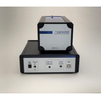
Introduction: Previous studies using a tissue-on-chip device quantified small extracellular vesicles (sEV) released from thyroid tissues, finding no differences in size or concentration, but differences in sEV content. To determine whether sEV release from tissue is reflected in the serum of the same patients [thyroid cancer (n = 6), Graves’ disease (n = 6), and benign thyroid (n = 6)], we employed the optical particle analysis instrument CX-300 (Paraytec, York, UK).
引言:此前使用组织芯片装置的研究对甲状腺组织释放的小细胞外囊泡(sEV)进行了定量,发现其大小或浓度没有差异,但 sEV 内容物存在差异。为确定组织释放的 sEV 是否反映在同一患者的血清中[甲状腺癌(n = 6)、Graves 病(n = 6)和良性甲状腺(n = 6)],我们采用了光学颗粒分析仪 CX-300(Paraytec,英国约克)。
Method: Serum was diluted 6.25- and 12.5-fold in phosphate buffered saline (PBS). Fifty microlitres of diluted serum was incubated with 2.5 µL fluorescein isothiocyanate (FITC)-conjugated anti-human CD63 antibody (BioLegend) for 60 min. Unbound antibody was removed using a ParaySelectTM column (Paraytec), which selectively retains labelled particles. Eluted particles were analysed using the CX-300 (three 60-second runs per sample).
方法:将血清用磷酸盐缓冲盐水(PBS)稀释 6.25 倍和 12.5 倍。将 50 微升稀释血清与 2.5 微升异硫氰酸荧光素(FITC)标记的抗人 CD63 抗体(BioLegend)孵育 60 分钟。使用 ParaySelectTM 柱(Paraytec)去除未结合的抗体,该柱可选择性保留标记颗粒。洗脱的颗粒使用 CX-300 进行分析(每个样品三次 60 秒运行)。
Results: One sample displayed high baseline fluorescence, preventing analysis. In the remaining samples, differential expression of CD63 was observed between pathologies., Serum from Graves’ disease patients contained significantly more CD63+ particles than serum from thyroid cancer patients (P < 0.05). Significant differences were also observed across all three patient groups.
结果:一个样品显示高基线荧光,无法分析。在其余样品中,观察到不同病理之间 CD63 的差异表达。Graves 病患者血清中含有的 CD63+ 颗粒显著多于甲状腺癌患者血清(P < 0.05)。在全部三个患者组之间也观察到显著差异。
Conclusion: The CX-300 fluorescence-based particle analysis system enabled quantification of CD63+ particles in serum. CD63 expression varied between thyroid pathologies, in contrast to earlier tissue-on-chip studies that used nanoparticle tracking analysis, which does not assess marker expression. Ongoing work aims to use a two-colour CX-300 system to investigate the expression of an additional tetraspanin marker, CD81.
结论:基于荧光的 CX-300 颗粒分析系统能够对血清中的 CD63+ 颗粒进行定量。CD63 表达在不同甲状腺病理之间发生变化,这与早期使用纳米颗粒跟踪分析的组织芯片研究相反,后者不评估标志物表达。正在进行的工作旨在使用双色 CX-300 系统研究另一种四跨膜蛋白标志物 CD81 的表达。